These are high-molecular organic compounds, biopolymers, built from 20 types of L-?-amino acid residues connected in a certain sequence into long chains. The molecular weight of proteins varies from 5 thousand to 1 million. The name “whites” was first given to the substance of bird eggs, which coagulates when heated into a white insoluble mass. The term was later extended to other substances with similar properties isolated from animals and plants.
Rice. 1. The most complex biopolymers are proteins. Their macromolecules consist of monomers, which are amino acids. Each amino acid has two functional groups: a carboxyl group and an amino group. All the diversity of proteins is created as a result of different combinations of 20 amino acids.
Proteins predominate over all other compounds present in living organisms, usually accounting for more than half of their dry weight. It is assumed that there are several billion individual proteins in nature (for example, more than 3 thousand different proteins are present in the E. coli bacterium alone).
Proteins play a key role in the life processes of any organism. Proteins include enzymes, with the participation of which all chemical transformations occur in the cell (metabolism); they control the action of genes; with their participation, the action of hormones is realized, transmembrane transport is carried out, including the generation of nerve impulses. They are an integral part of the immune system (immunoglobulins) and the coagulation system, form the basis of bone and connective tissue, and are involved in the transformation and utilization of energy.
History of protein research
The first attempts to isolate proteins were made back in the 18th century. By the beginning of the 19th century, the first works on the chemical study of proteins appeared. French scientists Joseph Louis Gay-Lussac and Louis Jacques Thénard tried to establish the elemental composition of proteins from different sources, which marked the beginning of systematic analytical studies, thanks to which it was concluded that all proteins are similar in the set of elements included in their composition. In 1836, the Dutch chemist G. J. Mulder proposed the first theory of the structure of protein substances, according to which all proteins have a certain hypothetical radical (C 40 H 62 N 10 O 12), associated in various proportions with sulfur and phosphorus atoms. He called this radical “protein” (from the Greek protein - first, main). Mulder's theory contributed to increasing interest in the study of proteins and improving the methods of protein chemistry. Techniques for isolating proteins by extraction with solutions of neutral salts were developed, and proteins were obtained in crystalline form for the first time (some plant proteins). To analyze proteins, they began to use their preliminary digestion with acids and alkalis.
At the same time, increasing attention began to be paid to the study of protein function. Jens Jakob Berzelius was the first to suggest in 1835 that they play the role of biocatalysts. Soon, proteolytic enzymes were discovered - pepsin (T. Schwann, 1836) and trypsin (L. Corvisart, 1856), which attracted attention to the physiology of digestion and the analysis of products formed during the breakdown of nutrients. Further studies of protein structure and work on the chemical synthesis of peptides resulted in the emergence of the peptide hypothesis, according to which all proteins are built from amino acids. By the end of the 19th century, most of the amino acids that make up proteins were studied.
At the beginning of the 20th century, the German chemist Emil Hermann Fischer was the first to use the methods of organic chemistry to study proteins and proved that proteins consist of β-amino acids linked to each other by an amide (peptide) bond. Later, thanks to the use of physicochemical methods of analysis, the molecular mass of many proteins was determined, the spherical shape of globular proteins was established, X-ray diffraction analysis of amino acids and peptides was carried out, and methods of chromatographic analysis were developed (see chromatography).
The first protein hormone was isolated (Frederick Grant Banting, John James Rickard McLeod, 1922), the presence of gamma globulins in antibodies was proven, and the enzymatic function of the muscle protein myosin was described (Vladimir Aleksandrovich Engelhardt, M. N. Lyubimova, 1939). For the first time, enzymes were obtained in crystalline form - urease (J.B. Saliner, 1926), pepsin (J.H. Nortron, 1929), lysozyme (E.P. Abraham, Robert Robinson, 1937).

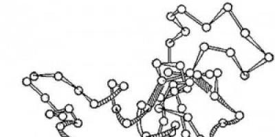
Rice. 2. Scheme of the three-dimensional structure of the enzyme lysozyme. Circles - amino acids; strands - peptide bonds; shaded rectangles are disulfide bonds. Spiralized and elongated sections of the polypeptide chain are visible.
In the 1950s, the three-level organization of protein molecules was proven - the presence of a primary, secondary and tertiary structure; created an automatic amino acid analyzer (Stanford Moore, William Howard Stein, 1950). In the 60s, attempts were made to chemically synthesize proteins (insulin, ribonuclease). X-ray diffraction analysis methods have been significantly improved; a device was created - a sequencer (P. Edman, G. Begg, 1967), which made it possible to determine the sequence of amino acids in a polypeptide chain. The consequence of this was the establishment of the structure of several hundred proteins from a variety of sources. Among them are proteolytic enzymes (pepsin, trypsin, chymotrypsin, subtilisin, carboxypeptidases), myoglobins, hemoglobins, cytochromes, lysozymes, immunoglobulins, histones, neurotoxins, viral envelope proteins, protein-peptide hormones. As a result, prerequisites emerged for solving pressing problems in enzymology, immunology, endocrinology and other areas of biological chemistry.
At the end of the 20th century, significant progress was made in studying the role of proteins in the matrix synthesis of biopolymers, understanding the mechanisms of their action in various life processes of organisms, and establishing the connection between their structure and function. The improvement of research methods and the emergence of new methods for separating proteins and peptides were of great importance.
The development of an effective method for analyzing the sequence of nucleotides in nucleic acids has made it possible to significantly simplify and speed up the determination of the amino acid sequence in proteins. This turned out to be possible because the order of amino acids in a protein is determined by the sequence of nucleotides in the gene encoding this protein (fragment). Consequently, knowing the arrangement of nucleotides in this gene and the genetic code, one can accurately predict in what order the amino acids are located in the polypeptide chain of a protein. Along with advances in the structural analysis of proteins, significant results have been achieved in the study of their spatial organization, mechanisms of formation and action of supramolecular complexes, including ribosomes and other cellular organelles, chromatin, viruses, etc.
Protein structure
Almost all proteins are built from 20 α-amino acids belonging to the L-series, and are the same in almost all organisms. Amino acids in proteins are connected to each other by a peptide bond -CO-NH-, which is formed by the carboxyl and -amino group of neighboring amino acid residues: two amino acids form a dipeptide in which the terminal carboxyl (-COOH) and amino group (H 2 N-) remain free, to to which new amino acids can be added to form a polypeptide chain.
The section of the chain on which the terminal H 2 N-group is located is called N-terminal, and the part opposite to it is called C-terminal. The huge variety of proteins is determined by the sequence of arrangement and the number of amino acid residues they contain. Although there is no clear distinction, short chains are usually called peptides or oligopeptides (from oligo...), and polypeptides (proteins) are usually understood as chains consisting of 50 or more. The most common proteins are those containing 100-400 amino acid residues, but there are also those whose molecules are formed by 1000 or more residues. Proteins can consist of several polypeptide chains. In such proteins, each polypeptide chain is called a subunit.
Spatial structure of proteins

Rice. 3. Protein in all organisms consists of 20 types of amino acids. Each protein is characterized by a certain assortment and quantitative ratio of amino acids. In protein molecules, amino acids are connected to each other by peptide bonds (- CO - NH -) in a linear sequence, constituting the so-called primary structure of the protein. Top line - free amino acids with side groups R1, R2, R3; bottom line - amino acids are connected by peptide bonds.
The polypeptide chain is capable of spontaneously forming and maintaining a special spatial structure. Based on the shape of protein molecules, proteins are divided into fibrillar and globular. In globular proteins, one or more polypeptide chains are folded into a compact spherical structure, or globule. Typically these proteins are highly soluble in water. These include almost all enzymes, blood transport proteins and many storage proteins. Fibrillar proteins are thread-like molecules held together by cross-links and form long fibers or layered structures. They have high mechanical strength, are insoluble in water and perform mainly structural and protective functions. Typical representatives of such proteins are hair and wool keratins, silk fibroin, and tendon collagen.
The order of covalently linked amino acids in a polypeptide chain is called the amino acid sequence, or the primary structure of proteins. The primary structure of each protein, encoded by the corresponding gene, is constant and carries all the information necessary for the formation of higher-level structures. The potential number of proteins that can be formed from 20 amino acids is practically unlimited.
As a result of the interaction of side groups of amino acid residues, individual relatively small sections of the polypeptide chain take on one or another conformation (type of folding), known as the secondary structure of proteins. Its most characteristic elements are the periodically repeating α-helix and β-structure. The secondary structure is very stable. Since it is largely determined by the amino acid sequence of the corresponding protein region, it becomes possible to predict it with a certain degree of probability. The term “?-helix” was introduced by the American biochemist, physicist and chemist Linus Carl Pauling, who described the arrangement of the polypeptide chain in the protein?-keratin in the form of a right-handed helix (the?-helix can be compared to a telephone cord). For each turn of such a helix in a protein there are 3.6 amino acid residues. This means that the -C=O group of one peptide bond forms a hydrogen bond with the -NH group of another peptide bond, four amino acid residues distant from the first one. On average, each α-helical region includes up to 15 amino acids, which corresponds to 3-4 turns of the helix. But in each individual protein, the length of the helix can differ greatly from this value. In cross section, the α-helix has the shape of a disk, from which the side chains of amino acids point outward.
Structure, or? -folded layer, can be formed by several sections of the polypeptide chain. These sections are stretched and laid parallel to each other, connected to each other by hydrogen bonds that occur between peptide bonds. They can be oriented in the same or opposite directions (the direction of movement along the polypeptide chain is usually considered to be from the N-terminus to the C-terminus). In the first case, the folded layer is called parallel, in the second - antiparallel. The latter is formed when the peptide chain makes a sharp turn back, forming a bend (?-bend). Are the amino acid side chains oriented perpendicular to the plane? -layer.
Relative content? -spiral sections and? -structures can vary widely among different proteins. There are proteins with a predominance of α-helices (about 75% of amino acids in myoglobin and hemoglobin), and the main type of chain folding in many fibrillar proteins (including silk fibroin, β-keratin) is α-helix. -structure. Regions of the polypeptide chain that cannot be classified into any of the above-described conformations are called connecting loops. Their structure is determined mainly by the interactions between the side chains of amino acids, and in the molecule of any protein it fits in a strictly defined way.
The tertiary structure is called spatial structure of globular proteins. But often this concept refers to the method of folding the polypeptide chain in space, characteristic of each specific protein. The tertiary structure is formed by the polypeptide chain of a protein spontaneously, apparently, along a certain coagulation path(s) with the preliminary formation of secondary structure elements. If the stability of the secondary structure is due to hydrogen bonds, then the tertiary structure is fixed by a diverse system of non-covalent interactions: hydrogen, ionic, intermolecular interactions, as well as hydrophobic contacts between the side chains of non-polar amino acid residues.
In some proteins, the tertiary structure is further stabilized by the formation of disulfide bonds (-S-S- bonds) between cysteine residues. As a rule, inside the protein globule there are side chains of hydrophobic amino acids assembled into the core (their transfer inside the protein globule is thermodynamically favorable), and on the periphery there are hydrophilic residues and some hydrophobic ones. The protein globule is surrounded by several hundred molecules of hydration water, which is necessary for the stability of the protein molecule and is often involved in its functioning. The tertiary structure is mobile, its individual sections can shift, which leads to conformational transitions that play a significant role in the interaction of the protein with other molecules.
Tertiary structure is the basis of the functional properties of a protein. It determines the formation of ensembles of functional groups in the protein - active centers and binding zones, gives them the necessary geometry, allows the creation of an internal environment, which is a prerequisite for the occurrence of many reactions, and ensures interaction with other proteins.
The tertiary structure of proteins clearly corresponds to its primary structure; there is probably an as yet undeciphered stereochemical code that determines the nature of protein folding. However, one and the same method of spatial arrangement usually corresponds not to a single primary structure, but to a whole family of structures in which only a small fraction (up to 20-30%) of amino acid residues may coincide, but in certain places in the chain the similarity of amino acid residues is preserved. The result is the formation of large families of proteins characterized by similar tertiary and more or less similar primary structure and, as a rule, common function. These are, for example, proteins of organisms of different species that have the same function and are evolutionarily related: myoglobins and hemoglobins, trypsin, chymotrypsin, elastase and other animal proteinases.

Rice. 4. As a result of the combination of several protein macromolecules with a tertiary structure, a quaternary protein structure is formed into a complex complex. An example of such complex proteins is hemoglobin, consisting of four macromolecules.
Often, especially in large proteins, the folding of a polypeptide chain occurs through the formation by individual sections of the chain of more or less autonomous elements of spatial structure - domains that can have functional autonomy, being responsible for one or another biological activity of the protein. Thus, the N-terminal domains of blood coagulation proteins ensure their attachment to the cell membrane.
There are many proteins whose molecules are an ensemble of globules (subunits) held together by hydrophobic interactions, hydrogen or ionic bonds. Such complexes are called oligomeric, multimeric or subunit proteins. The arrangement of subunits in a functionally active protein complex is called the quaternary structure of the protein. Some proteins are capable of forming structures of higher orders, for example, multienzyme complexes, extended structures (bacteriophage coat proteins), supramolecular complexes that function as a single whole (for example, ribosomes or components of the mitochondrial respiratory chain).
The quaternary structure allows the creation of molecules with unusual geometries. Thus, ferritin, formed by 24 subunits, has an internal cavity, thanks to which the protein manages to bind up to 3000 iron ions. In addition, the quaternary structure allows several different functions to be performed in one molecule. Tryptophan synthetase combines enzymes responsible for several successive stages of the synthesis of the amino acid tryptophan.
Methods for studying protein structure
The primary structure of proteins determines all other levels of organization of the protein molecule. Therefore, when studying the biological function of various proteins, knowledge of this structure is important. The first protein for which the amino acid sequence was established was the pancreatic hormone, insulin. This work, which took 11 years, was carried out by the English biochemist Frederick Sanger (1954). He determined the location of 51 amino acids in the hormone molecule and showed that it consists of 2 chains connected by disulfide bonds. Later, most of the work on establishing the primary structure of proteins was automated.
With the development of genetic engineering methods, it became possible to further accelerate this process by determining the primary structure of proteins in accordance with the results of analysis of the nucleotide sequence in the genes encoding these proteins. The secondary and tertiary structure of proteins is studied using quite complex physical methods, for example, circular dichroism or X-ray diffraction analysis of protein crystals. The tertiary structure was first established by the English biochemist John Cowdery Kendrew (1957) for the muscle protein myoglobin.

Rice. 5. Model of the myoglobin molecule (spatial configuration of the molecule)
Denaturation of proteins
Relatively weak bonds responsible for stabilizing the secondary, tertiary and quaternary structures of the protein are easily destroyed, which is accompanied by a loss of its biological activity. The destruction of the original (native) protein structure, called denaturation, occurs in the presence of acids and bases, with heating, changes in ionic strength and other influences. As a rule, denatured proteins are poorly or not at all soluble in water. With a short-term effect and rapid elimination of denaturing factors, protein renaturation is possible with complete or partial restoration of the original structure and biological properties.
Protein classification
The complexity of the structure of protein molecules and the extreme variety of functions they perform make it difficult to create a unified and clear classification of them, although attempts to do this have been made repeatedly since the end of the 19th century. Based on their chemical composition, proteins are divided into simple and complex (sometimes called proteids). The molecules of the former consist only of amino acids. In addition to the polypeptide chain itself, complex proteins contain non-protein components represented by carbohydrates (glycoproteins), lipids (lipoproteins), nucleic acids (nucleoproteins), metal ions (metalloproteins), phosphate group (phosphoproteins), pigments (chromoproteins), etc. .
Depending on the functions they perform, several classes of proteins are distinguished. The most diverse and most specialized class consists of proteins with a catalytic function - enzymes that have the ability to accelerate chemical reactions occurring in living organisms. In this capacity, proteins participate in all processes of synthesis and breakdown of various compounds during metabolism, in the biosynthesis of proteins and nucleic acids, regulation of cell development and differentiation. Transport proteins have the ability to selectively bind fatty acids, hormones and other organic and inorganic compounds and ions, and then transport them with current to the desired location (for example, hemoglobin is involved in the transfer of oxygen from the lungs to all cells of the body). Transport proteins also carry out active transport of ions, lipids, sugars and amino acids across biological membranes.
Structural proteins perform a supporting or protective function; they participate in the formation of the cell skeleton. The most common among them are collagen of connective tissue, keratin, nails and feathers, elastin of vascular cells and many others. In combination with lipids, they are the structural basis of cellular and intracellular membranes.
A number of proteins perform a protective function. For example, immunoglobulins (antibodies) of vertebrates, having the ability to bind foreign pathogenic microorganisms and substances, neutralize their pathogenic effects on the body and prevent cell proliferation. Fibrinogen and thrombin are involved in the blood clotting process. Many protein substances secreted by bacteria, as well as components of some invertebrates, are classified as toxins.
Some proteins (regulatory) are involved in the regulation of the physiological activity of the body as a whole, individual organs, cells or processes. They control gene transcription and protein synthesis; these include peptide-protein hormones secreted by endocrine glands. Seed storage proteins provide nutrients for the initial stages of embryo development. These also include casein, egg white albumin (ovalbumin) and many others. Thanks to proteins, muscle cells acquire the ability to contract and ultimately provide movement to the body. Examples of such contractile proteins are skeletal muscle actin and myosin, as well as tubulin, which are components of the cilia and flagella of unicellular organisms; They also ensure the divergence of chromosomes during cell division.
Receptor proteins are the target of hormones and other biologically active compounds. With their help, the cell perceives information about the state of the external environment. They play an important role in the transmission of nervous excitation and in oriented cell movement (chemotaxis). The transformation and utilization of energy entering the body, as well as energy, also occurs with the participation of proteins of the bioenergy system (for example, the visual pigment rhodopsin, cytochromes of the respiratory chain). There are also many proteins with other, sometimes rather unusual functions (for example, the plasma of some Antarctic fish contains proteins that have antifreeze properties).
Protein biosynthesis
All information about the structure of a particular protein is “stored” in the corresponding genes in the form of a sequence of nucleotides and is implemented in the process of template synthesis. First, information is transferred (read) from the DNA molecule to messenger RNA (mRNA) using the enzyme DNA-dependent RNA polymerase, and then in the ribosome on the mRNA, as on a matrix in accordance with the genetic code, with the participation of transport RNAs delivering amino acids, the formation occurs polypeptide chain.
The synthesized polypeptide chains emerging from the ribosome, spontaneously folding, take on the conformation characteristic of the protein and can be subject to post-translational modification. The side chains of individual amino acids can undergo modifications (hydroxylation, phosphorylation, etc.). That is why, for example, hydroxyproline and hydroxylysine are found in collagen (see). The modification may also be accompanied by the rupture of polypeptide bonds. In this way, for example, the formation of an active insulin molecule occurs, consisting of two chains connected by disulfide bonds.

Rice. 6. General scheme of protein biosynthesis.
The importance of proteins in nutrition
Proteins are the most important components of animal and human food. The nutritional value of proteins is determined by their content of essential amino acids, which are not produced in the body itself. In this regard, plant proteins are less valuable than animal proteins: they are poorer in lysine, methionine and tryptophan, and are more difficult to digest in the gastrointestinal tract. The lack of essential amino acids in food leads to severe disorders of nitrogen metabolism.
Proteins are broken down into free amino acids, which, after absorption in the intestine, enter and are distributed to all cells. Some of them break down into simple compounds with the release of energy, used for various needs by the cell, and some go to the synthesis of new proteins characteristic of a given organism. (R. A. Matveeva, Encyclopedia Cyril and Methodius)
Enumeration of proteins
- amyloid - amyloid;
- anionic - anionic;
- antivirus - antiviral;
- autoimmune - autoimmune;
- autologous - autologic;
- bacterial - bacterial;
- Bence Jones protein;
- virus-induced - virus induced;
- viral - virus;
- viral nonstructural - virus nonstructural;
- viral structural - virus structural;
- virus-specific - virus specific;
- high molecular weight - high molecular weight;
- heme-containing - heme;
- heterologous - foreign;
- hybrid - hybrid;
- glycosylated - glycated;
- globular - globular;
- denatured - denatured;
- iron-containing - iron;
- yolk - yolk;
- animal protein - animal protein;
- protective - defensive;
- immune - immune;
- immunogenic - immunologically relevant;
- calcium binding;
- sour - acidic;
- corpuscular - corpuscular;
- membrane - membrane;
- myeloma - myeloma;
- microsomal - microsomal;
- milk protein - milk protein;
- monoclonal - monoclonal immunoglobulin;
- muscle protein - muscle protein;
- native - native;
- nonhistone - nonhistone;
- defective - partial;
- insoluble - insoluble;
- indigestible - insoluble;
- non-enzymatic - nonenzyme;
- low molecular weight - low molecular weight;
- new protein - new protein;
- general - whole;
- oncogenic - oncoprotein;
- main phase protein - anionic;
- protein of acute phase (inflammation) - protein of acute phase;
- food - food;
- blood plasma protein - plasma protein;
- placental - placenta;
- uncoupling - uncoupling;
- protein of regenerating nerve;
- regulatory - regulatory;
- recombination - recombinant;
- receptor - receptor;
- ribosomal - ribosomal;
- binding - binding;
- secretory protein - secretory protein;
- C-reactive - C-reactive;
- whey protein - whey protein, lactoprotein;
- tissue - tissue;
- toxic - toxic;
- chimeric - chimeric;
- whole - whole;
- cytosolic - cytosolic;
- alkaline protein - anionic protein;
- exogenous - exogenous;
- endogenous - endogenous protein.
Read more about proteins in the literature:
- Volkenshtein M.V., Molecules and, M., 1965, ch. 3 - 5;
- Gaurowitz F., Chemistry and functions of proteins, trans. from English, Moscow, 1965;
- Sissakyan N. M. and Gladilin K. L., Biochemical aspects of protein synthesis, in the book: Advances in biological chemistry, vol. 7, M., 1965, p. 3;
- Stepanov V. M. Molecular biology. Structure and function of proteins. M., 1996;
- Shamin A. N., Development of protein chemistry, M., 1966;
- Proteins and peptides. M., 1995-2000. T. 1-3;
- Biosynthesis of protein and nucleic acids, ed. A. S. Spirina, M., 1965;
- Introduction to molecular biology, trans. from English, M., 1967
- Molecules and cells. [Sat. Art.], trans. from English, M., 1966, p. 7 - 27, 94 - 106;
- Fundamentals of biochemistry: Translation from English M., 1981. T. 1;
- The protein problem. M., 1995. T. 1-5;
- The Proteins. New York, 1975-79. 3 ed. V. 1-4.
Find something else interesting:
Squirrels- high molecular weight organic compounds consisting of α-amino acid residues.
IN protein composition includes carbon, hydrogen, nitrogen, oxygen, sulfur. Some proteins form complexes with other molecules containing phosphorus, iron, zinc and copper.
Proteins have a large molecular weight: egg albumin - 36,000, hemoglobin - 152,000, myosin - 500,000. For comparison: the molecular weight of alcohol is 46, acetic acid - 60, benzene - 78.
Amino acid composition of proteins
Squirrels- non-periodic polymers, the monomers of which are α-amino acids. Typically, 20 types of α-amino acids are called protein monomers, although over 170 of them are found in cells and tissues.
Depending on whether amino acids can be synthesized in the body of humans and other animals, they are distinguished: nonessential amino acids- can be synthesized; essential amino acids- cannot be synthesized. Essential amino acids must be supplied to the body through food. Plants synthesize all types of amino acids.
Depending on the amino acid composition, proteins are: complete- contain the entire set of amino acids; defective- some amino acids are missing in their composition. If proteins consist only of amino acids, they are called simple. If proteins contain, in addition to amino acids, a non-amino acid component (prosthetic group), they are called complex. The prosthetic group can be represented by metals (metalloproteins), carbohydrates (glycoproteins), lipids (lipoproteins), nucleic acids (nucleoproteins).
All amino acids contain: 1) carboxyl group (-COOH), 2) amino group (-NH 2), 3) radical or R-group (the rest of the molecule). The structure of the radical is different for different types of amino acids. Depending on the number of amino groups and carboxyl groups included in the composition of amino acids, they are distinguished: neutral amino acids having one carboxyl group and one amino group; basic amino acids having more than one amino group; acidic amino acids having more than one carboxyl group.
Amino acids are amphoteric compounds, since in solution they can act as both acids and bases. In aqueous solutions, amino acids exist in different ionic forms.
Peptide bond
Peptides- organic substances consisting of amino acid residues connected by peptide bonds.
The formation of peptides occurs as a result of the condensation reaction of amino acids. When the amino group of one amino acid interacts with the carboxyl group of another, a covalent nitrogen-carbon bond occurs between them, which is called peptide. Depending on the number of amino acid residues included in the peptide, there are dipeptides, tripeptides, tetrapeptides etc. The formation of a peptide bond can be repeated many times. This leads to the formation polypeptides. At one end of the peptide there is a free amino group (called the N-terminus), and at the other there is a free carboxyl group (called the C-terminus).

Spatial organization of protein molecules
The performance of certain specific functions by proteins depends on the spatial configuration of their molecules; in addition, it is energetically unfavorable for the cell to keep proteins in an unfolded form, in the form of a chain, therefore polypeptide chains undergo folding, acquiring a certain three-dimensional structure, or conformation. There are 4 levels spatial organization of proteins.
Primary protein structure- the sequence of arrangement of amino acid residues in the polypeptide chain that makes up the protein molecule. The bond between amino acids is a peptide bond.

If a protein molecule consists of only 10 amino acid residues, then the number of theoretically possible variants of protein molecules that differ in the order of alternation of amino acids is 10 20. Having 20 amino acids, you can make even more diverse combinations from them. About ten thousand different proteins have been found in the human body, which differ both from each other and from the proteins of other organisms.
It is the primary structure of the protein molecule that determines the properties of the protein molecules and its spatial configuration. Replacing just one amino acid with another in a polypeptide chain leads to a change in the properties and functions of the protein. For example, replacing the sixth glutamic amino acid in the β-subunit of hemoglobin with valine leads to the fact that the hemoglobin molecule as a whole cannot perform its main function - oxygen transport; In such cases, the person develops a disease called sickle cell anemia.
Secondary structure- ordered folding of the polypeptide chain into a spiral (looks like an extended spring). The turns of the helix are strengthened by hydrogen bonds that arise between carboxyl groups and amino groups. Almost all CO and NH groups take part in the formation of hydrogen bonds. They are weaker than peptide ones, but, repeated many times, impart stability and rigidity to this configuration. At the level of secondary structure, there are proteins: fibroin (silk, spider web), keratin (hair, nails), collagen (tendons).


Tertiary structure- packing of polypeptide chains into globules, resulting from the formation of chemical bonds (hydrogen, ionic, disulfide) and the establishment of hydrophobic interactions between the radicals of amino acid residues. The main role in the formation of the tertiary structure is played by hydrophilic-hydrophobic interactions. In aqueous solutions, hydrophobic radicals tend to hide from water, grouping inside the globule, while hydrophilic radicals, as a result of hydration (interaction with water dipoles), tend to appear on the surface of the molecule. In some proteins, the tertiary structure is stabilized by disulfide covalent bonds formed between the sulfur atoms of two cysteine residues. At the tertiary structure level there are enzymes, antibodies, and some hormones.

Quaternary structure characteristic of complex proteins whose molecules are formed by two or more globules. The subunits are held in the molecule by ionic, hydrophobic, and electrostatic interactions. Sometimes, during the formation of a quaternary structure, disulfide bonds occur between subunits. The most studied protein with a quaternary structure is hemoglobin. It is formed by two α-subunits (141 amino acid residues) and two β-subunits (146 amino acid residues). Associated with each subunit is a heme molecule containing iron.
If for some reason the spatial conformation of proteins deviates from normal, the protein cannot perform its functions. For example, the cause of “mad cow disease” (spongiform encephalopathy) is the abnormal conformation of prions, the surface proteins of nerve cells.
Properties of proteins
The amino acid composition and structure of the protein molecule determine it properties. Proteins combine basic and acidic properties, determined by amino acid radicals: the more acidic amino acids in a protein, the more pronounced its acidic properties. The ability to donate and add H + is determined buffering properties of proteins; One of the most powerful buffers is hemoglobin in red blood cells, which maintains blood pH at a constant level. There are soluble proteins (fibrinogen), and there are insoluble proteins that perform mechanical functions (fibroin, keratin, collagen). There are proteins that are chemically active (enzymes), there are chemically inactive proteins that are resistant to various environmental conditions and those that are extremely unstable.
External factors (heat, ultraviolet radiation, heavy metals and their salts, pH changes, radiation, dehydration)
can cause disruption of the structural organization of the protein molecule. The process of loss of the three-dimensional conformation inherent in a given protein molecule is called denaturation. The cause of denaturation is the breaking of bonds that stabilize a certain protein structure. Initially, the weakest ties are broken, and as conditions become stricter, even stronger ones are broken. Therefore, first the quaternary, then the tertiary and secondary structures are lost. A change in spatial configuration leads to a change in the properties of the protein and, as a result, makes it impossible for the protein to perform its inherent biological functions. If denaturation is not accompanied by destruction of the primary structure, then it may be reversible, in this case, self-recovery of the conformation characteristic of the protein occurs. For example, membrane receptor proteins undergo such denaturation. The process of restoring protein structure after denaturation is called renaturation. If restoration of the spatial configuration of the protein is impossible, then denaturation is called irreversible.
Functions of proteins
| Function | Examples and explanations |
|---|---|
| Construction | Proteins are involved in the formation of cellular and extracellular structures: they are part of cell membranes (lipoproteins, glycoproteins), hair (keratin), tendons (collagen), etc. |
| Transport | The blood protein hemoglobin attaches oxygen and transports it from the lungs to all tissues and organs, and from them transfers carbon dioxide to the lungs; The composition of cell membranes includes special proteins that ensure the active and strictly selective transfer of certain substances and ions from the cell to the external environment and back. |
| Regulatory | Protein hormones take part in the regulation of metabolic processes. For example, the hormone insulin regulates blood glucose levels, promotes glycogen synthesis, and increases the formation of fats from carbohydrates. |
| Protective | In response to the penetration of foreign proteins or microorganisms (antigens) into the body, special proteins are formed - antibodies that can bind and neutralize them. Fibrin, formed from fibrinogen, helps stop bleeding. |
| Motor | The contractile proteins actin and myosin provide muscle contraction in multicellular animals. |
| Signal | Built into the surface membrane of the cell are protein molecules that are capable of changing their tertiary structure in response to environmental factors, thus receiving signals from the external environment and transmitting commands to the cell. |
| Storage | In the body of animals, proteins, as a rule, are not stored, with the exception of egg albumin and milk casein. But thanks to proteins, some substances can be stored in the body; for example, during the breakdown of hemoglobin, iron is not removed from the body, but is stored, forming a complex with the protein ferritin. |
| Energy | When 1 g of protein breaks down into final products, 17.6 kJ is released. First, proteins break down into amino acids, and then into the final products - water, carbon dioxide and ammonia. However, proteins are used as a source of energy only when other sources (carbohydrates and fats) are used up. |
| Catalytic | One of the most important functions of proteins. Provided by proteins - enzymes that accelerate biochemical reactions occurring in cells. For example, ribulose biphosphate carboxylase catalyzes the fixation of CO 2 during photosynthesis. |
Enzymes
Enzymes, or enzymes, are a special class of proteins that are biological catalysts. Thanks to enzymes, biochemical reactions occur at tremendous speed. The speed of enzymatic reactions is tens of thousands of times (and sometimes millions) higher than the speed of reactions occurring with the participation of inorganic catalysts. The substance on which the enzyme acts is called substrate.
Enzymes are globular proteins, structural features enzymes can be divided into two groups: simple and complex. Simple enzymes are simple proteins, i.e. consist only of amino acids. Complex enzymes are complex proteins, i.e. In addition to the protein part, they contain a group of non-protein nature - cofactor. Some enzymes use vitamins as cofactors. The enzyme molecule contains a special part called the active center. Active center- a small section of the enzyme (from three to twelve amino acid residues), where the binding of the substrate or substrates occurs to form an enzyme-substrate complex. Upon completion of the reaction, the enzyme-substrate complex breaks down into the enzyme and the reaction product(s). Some enzymes have (except active) allosteric centers- areas to which enzyme speed regulators are attached ( allosteric enzymes).

Reactions of enzymatic catalysis are characterized by: 1) high efficiency, 2) strict selectivity and direction of action, 3) substrate specificity, 4) fine and precise regulation. The substrate and reaction specificity of enzymatic catalysis reactions are explained by the hypotheses of E. Fischer (1890) and D. Koshland (1959).
E. Fisher (key-lock hypothesis) suggested that the spatial configurations of the active center of the enzyme and the substrate must correspond exactly to each other. The substrate is compared to the “key”, the enzyme to the “lock”.
D. Koshland (hand-glove hypothesis) suggested that the spatial correspondence between the structure of the substrate and the active center of the enzyme is created only at the moment of their interaction with each other. This hypothesis is also called induced correspondence hypothesis.
The rate of enzymatic reactions depends on: 1) temperature, 2) enzyme concentration, 3) substrate concentration, 4) pH. It should be emphasized that since enzymes are proteins, their activity is highest under physiologically normal conditions.
Most enzymes can only work at temperatures between 0 and 40°C. Within these limits, the reaction rate increases approximately 2 times with every 10 °C increase in temperature. At temperatures above 40 °C, the protein undergoes denaturation and enzyme activity decreases. At temperatures close to freezing, enzymes are inactivated.
As the amount of substrate increases, the rate of the enzymatic reaction increases until the number of substrate molecules equals the number of enzyme molecules. With a further increase in the amount of substrate, the speed will not increase, since the active centers of the enzyme are saturated. An increase in enzyme concentration leads to increased catalytic activity, since a larger number of substrate molecules undergo transformations per unit time.

For each enzyme, there is an optimal pH value at which it exhibits maximum activity (pepsin - 2.0, salivary amylase - 6.8, pancreatic lipase - 9.0). At higher or lower pH values, enzyme activity decreases. With sudden changes in pH, the enzyme denatures.
The speed of allosteric enzymes is regulated by substances that attach to allosteric centers. If these substances speed up a reaction, they are called activators, if they slow down - inhibitors.
Classification of enzymes
According to the type of chemical transformations they catalyze, enzymes are divided into 6 classes:
- oxireductases(transfer of hydrogen, oxygen or electron atoms from one substance to another - dehydrogenase),
- transferases(transfer of methyl, acyl, phosphate or amino group from one substance to another - transaminase),
- hydrolases(hydrolysis reactions in which two products are formed from the substrate - amylase, lipase),
- lyases(non-hydrolytic addition to the substrate or detachment of a group of atoms from it, in which case C-C, C-N, C-O, C-S bonds can be broken - decarboxylase),
- isomerases(intramolecular rearrangement - isomerase),
- ligases(the connection of two molecules as a result of the formation of C-C, C-N, C-O, C-S bonds - synthetase).
Classes are in turn subdivided into subclasses and subsubclasses. In the current international classification, each enzyme has a specific code, consisting of four numbers separated by dots. The first number is the class, the second is the subclass, the third is the subsubclass, the fourth is the serial number of the enzyme in this subclass, for example, the arginase code is 3.5.3.1.
Go to lectures No. 2"Structure and functions of carbohydrates and lipids"
Go to lectures No. 4"Structure and functions of ATP nucleic acids"
In the first half of the 19th century. many chemists, and among them primarily J. von Liebig, gradually came to the conclusion that proteins represent a special class of nitrogenous compounds. The name "proteins" (from the Greek.
protos first) was proposed in 1840 by the Dutch chemist G. Mulder. PHYSICAL PROPERTIES Proteins are white in the solid state, but colorless in solution, unless they carry some kind of chromophore (colored) group, such as hemoglobin. The solubility in water varies greatly among different proteins. It also changes depending on the pH and the concentration of salts in the solution, so it is possible to select conditions under which one protein will selectively precipitate in the presence of other proteins. This "salting out" method is widely used to isolate and purify proteins. The purified protein often precipitates out of solution as crystals.Compared to other compounds, the molecular weight of proteins is very large, ranging from several thousand to many millions of daltons. Therefore, during ultracentrifugation, proteins are sedimented, and at different rates. Due to the presence of positively and negatively charged groups in protein molecules, they move at different speeds and in an electric field. This is the basis of electrophoresis, a method used to isolate individual proteins from complex mixtures. Proteins are also purified by chromatography.
CHEMICAL PROPERTIES Structure. Proteins are polymers, i.e. molecules built like chains from repeating monomer units, or subunits, the role of which they play a -amino acids. General formula of amino acids where R a hydrogen atom or some organic group.A protein molecule (polypeptide chain) can consist of only a relatively small number of amino acids or several thousand monomer units. The combination of amino acids in a chain is possible because each of them has two different chemical groups: an amino group with basic properties,
NH 2 , and an acidic carboxyl group, COOH. Both of these groups are affiliated with a -carbon atom. The carboxyl group of one amino acid can form an amide (peptide) bond with the amino group of another amino acid: After two amino acids have been linked in this way, the chain can be extended by adding a third to the second amino acid, and so on. As can be seen from the above equation, when a peptide bond is formed, a water molecule is released. In the presence of acids, alkalis or proteolytic enzymes, the reaction proceeds in the opposite direction: the polypeptide chain is split into amino acids with the addition of water. This reaction is called hydrolysis. Hydrolysis occurs spontaneously, and energy is required to connect amino acids into a polypeptide chain.
 A carboxyl group and an amide group (or a similar imide group in the case of the amino acid proline) are present in all amino acids, but the differences between amino acids are determined by the nature of the group, or “side chain,” which is indicated above by the letter
R . The role of the side chain can be played by one hydrogen atom, as in the amino acid glycine, or by some bulky group, as in histidine and tryptophan. Some side chains are chemically inert, while others are markedly reactive.Many thousands of different amino acids can be synthesized, and many different amino acids occur in nature, but only 20 types of amino acids are used for protein synthesis: alanine, arginine, asparagine, aspartic acid, valine, histidine, glycine, glutamine, glutamic acid, isoleucine, leucine, lysine , methionine, proline, serine, tyrosine, threonine, tryptophan, phenylalanine and cysteine (in proteins, cysteine may be present as a dimer
cystine). True, some proteins contain other amino acids in addition to the regularly occurring twenty, but they are formed as a result of modification of one of the twenty listed after it has been included in the protein.Optical activity. All amino acids, with the exception of glycine, have a The -carbon atom has four different groups attached to it. From the point of view of geometry, four different groups can be attached in two ways, and accordingly there are two possible configurations, or two isomers, related to each other as an object is to its mirror image, i.e. like the left hand to the right. One configuration is called left, or left-handed ( L ), and the other right, or dextrorotatory ( D ), since two such isomers differ in the direction of rotation of the plane of polarized light. Found only in proteins L -amino acids (the exception is glycine; it can be represented in only one form, since two of its four groups are the same), and all of them are optically active (since there is only one isomer). D -amino acids are rare in nature; they are found in some antibiotics and the cell wall of bacteria.Amino acid sequence. Amino acids in a polypeptide chain are not arranged randomly, but in a certain fixed order, and it is this order that determines the functions and properties of the protein. By varying the order of the 20 types of amino acids, you can create a huge number of different proteins, just as you can create many different texts from the letters of the alphabet.In the past, determining the amino acid sequence of a protein often took several years. Direct determination is still quite a labor-intensive task, although devices have been created that allow it to be carried out automatically. It is usually easier to determine the nucleotide sequence of the corresponding gene and deduce the amino acid sequence of the protein from it. To date, the amino acid sequences of many hundreds of proteins have already been determined. The functions of the deciphered proteins are usually known, and this helps to imagine the possible functions of similar proteins formed, for example, in malignant neoplasms.
Complex proteins. Proteins consisting of only amino acids are called simple. Often, however, a metal atom or some chemical compound that is not an amino acid is attached to the polypeptide chain. Such proteins are called complex. An example is hemoglobin: it contains iron porphyrin, which determines its red color and allows it to act as an oxygen carrier.The names of most complex proteins indicate the nature of the attached groups: glycoproteins contain sugars, lipoproteins contain fats. If the catalytic activity of an enzyme depends on the attached group, then it is called a prosthetic group. Often a vitamin plays the role of a prosthetic group or is part of one. Vitamin A, for example, attached to one of the proteins in the retina, determines its sensitivity to light.
Tertiary structure. What is important is not so much the amino acid sequence of the protein itself (the primary structure), but the way it is laid out in space. Along the entire length of the polypeptide chain, hydrogen ions form regular hydrogen bonds, which give it the shape of a helix or layer (secondary structure). From the combination of such helices and layers, a compact form of the next order emerges: the tertiary structure of the protein. Around the bonds holding the monomer units of the chain, rotations at small angles are possible. Therefore, from a purely geometric point of view, the number of possible configurations for any polypeptide chain is infinitely large. In reality, each protein normally exists in only one configuration, determined by its amino acid sequence. This structure is not rigid, it is as if « breathes” fluctuates around a certain average configuration. The circuit is folded into a configuration in which free energy (the ability to produce work) is minimal, just as a released spring compresses only to a state corresponding to the minimum free energy. Often one part of the chain is rigidly linked to another by disulfide ( SS) bonds between two cysteine residues. This is partly why cysteine plays a particularly important role among amino acids.The complexity of the structure of proteins is so great that it is not yet possible to calculate the tertiary structure of a protein, even if its amino acid sequence is known. But if it is possible to obtain protein crystals, then its tertiary structure can be determined by X-ray diffraction.
In structural, contractile and some other proteins, the chains are elongated and several slightly folded chains lying nearby form fibrils; the fibrils, in turn, fold into larger formations of fibers. However, most proteins in solution have a globular shape: the chains are coiled in a globule, like yarn in a ball. Free energy with this configuration is minimal, since hydrophobic (“water-repelling”) amino acids are hidden inside the globule, and hydrophilic (“water-attracting”) amino acids are on its surface.
Many proteins are complexes of several polypeptide chains. This structure is called the quaternary structure of the protein. The hemoglobin molecule, for example, consists of four subunits, each of which is a globular protein.
Structural proteins, due to their linear configuration, form fibers that have a very high tensile strength, while the globular configuration allows the proteins to enter into specific interactions with other compounds. On the surface of the globule, when the chains are correctly laid out, cavities of a certain shape appear in which reactive chemical groups are located. If a given protein is an enzyme, then another, usually smaller, molecule of some substance enters such a cavity, just as a key enters a lock; in this case, the configuration of the electron cloud of the molecule changes under the influence of the chemical groups located in the cavity, and this forces it to react in a certain way. In this way, the enzyme catalyzes the reaction. Antibody molecules also have cavities in which various foreign substances bind and are thereby rendered harmless. The “lock and key” model, which explains the interaction of proteins with other compounds, allows us to understand the specificity of enzymes and antibodies, i.e. their ability to react only with certain compounds.
Proteins in different types of organisms. Proteins that perform the same function in different species of plants and animals and therefore bear the same name also have a similar configuration. They, however, differ somewhat in their amino acid sequence. As species diverge from a common ancestor, some amino acids at certain positions are replaced by mutations by others. Harmful mutations that cause hereditary diseases are eliminated by natural selection, but beneficial or at least neutral ones may persist. The closer two biological species are to each other, the less differences are found in their proteins.Some proteins change relatively quickly, others are very conserved. The latter includes, for example, cytochrome With a respiratory enzyme found in most living organisms. In humans and chimpanzees, its amino acid sequences are identical, and in cytochrome With In wheat, only 38% of the amino acids were different. Even comparing humans and bacteria, the similarity of cytochromes With(the differences affect 65% of the amino acids here) can still be seen, although the common ancestor of bacteria and humans lived on Earth about two billion years ago. Nowadays, comparison of amino acid sequences is often used to construct a phylogenetic (family) tree, reflecting the evolutionary relationships between different organisms.
Denaturation. The synthesized protein molecule, folding, acquires its characteristic configuration. This configuration, however, can be destroyed by heating, by changing pH, by exposure to organic solvents, and even by simply shaking the solution until bubbles appear on its surface. A protein modified in this way is called denatured; it loses its biological activity and usually becomes insoluble. Well-known examples of denatured protein are boiled eggs or whipped cream. Small proteins containing only about a hundred amino acids are capable of renaturation, i.e. reacquire the original configuration. But most proteins simply turn into a mass of tangled polypeptide chains and do not restore their previous configuration.One of the main difficulties in isolating active proteins is their extreme sensitivity to denaturation. This property of proteins finds useful application in food preservation: high temperature irreversibly denatures the enzymes of microorganisms, and the microorganisms die.
PROTEIN SYNTHESIS To synthesize protein, a living organism must have a system of enzymes capable of joining one amino acid to another. A source of information is also needed to determine which amino acids should be combined. Since there are thousands of types of proteins in the body and each of them consists on average of several hundred amino acids, the information required must be truly enormous. It is stored (similar to how a recording is stored on a magnetic tape) in the nucleic acid molecules that make up genes. Cm . also HEREDITARY; NUCLEIC ACIDS.Enzyme activation. A polypeptide chain synthesized from amino acids is not always a protein in its final form. Many enzymes are synthesized first as inactive precursors and become active only after another enzyme removes several amino acids at one end of the chain. Some of the digestive enzymes, such as trypsin, are synthesized in this inactive form; these enzymes are activated in the digestive tract as a result of the removal of the terminal fragment of the chain. The hormone insulin, the molecule of which in its active form consists of two short chains, is synthesized in the form of one chain, the so-called. proinsulin. The middle part of this chain is then removed, and the remaining fragments bind together to form the active hormone molecule. Complex proteins are formed only after a specific chemical group is attached to the protein, and this attachment often also requires an enzyme.Metabolic circulation. After feeding an animal amino acids labeled with radioactive isotopes of carbon, nitrogen or hydrogen, the label is quickly incorporated into its proteins. If labeled amino acids stop entering the body, the amount of label in proteins begins to decrease. These experiments show that the resulting proteins are not retained in the body until the end of life. All of them, with few exceptions, are in a dynamic state, constantly breaking down into amino acids and then being synthesized again.Some proteins break down when cells die and are destroyed. This happens all the time, for example, with red blood cells and epithelial cells lining the inner surface of the intestine. In addition, the breakdown and resynthesis of proteins also occurs in living cells. Oddly enough, less is known about the breakdown of proteins than about their synthesis. It is clear, however, that the breakdown involves proteolytic enzymes similar to those that break down proteins into amino acids in the digestive tract.
The half-life of different proteins varies from several hours to many months. The only exception is the collagen molecule. Once formed, they remain stable and are not renewed or replaced. Over time, however, some of their properties change, in particular elasticity, and since they are not renewed, this results in certain age-related changes, such as the appearance of wrinkles on the skin.
Synthetic proteins. Chemists have long learned to polymerize amino acids, but the amino acids are combined in a disorderly manner, so that the products of such polymerization bear little resemblance to natural ones. True, it is possible to combine amino acids in a given order, which makes it possible to obtain some biologically active proteins, in particular insulin. The process is quite complicated, and in this way it is possible to obtain only those proteins whose molecules contain about a hundred amino acids. It is preferable instead to synthesize or isolate the nucleotide sequence of a gene corresponding to the desired amino acid sequence, and then introduce this gene into a bacterium, which will produce large quantities of the desired product by replication. This method, however, also has its drawbacks. Cm . also GENETIC ENGINEERING. PROTEIN AND NUTRITION When proteins in the body are broken down into amino acids, these amino acids can be used again to synthesize proteins. At the same time, the amino acids themselves are subject to breakdown, so they are not completely reutilized. It is also clear that during growth, pregnancy and wound healing, protein synthesis must exceed breakdown. The body continuously loses some proteins; These are the proteins of hair, nails and the surface layer of skin. Therefore, in order to synthesize proteins, each organism must receive amino acids from food. Green plants synthesize from CO 2 , water and ammonia or nitrates are all 20 amino acids found in proteins. Many bacteria are also capable of synthesizing amino acids in the presence of sugar (or some equivalent) and fixed nitrogen, but sugar is ultimately supplied by green plants. Animals have a limited ability to synthesize amino acids; they obtain amino acids by eating green plants or other animals. In the digestive tract, absorbed proteins are broken down into amino acids, the latter are absorbed, and from them proteins characteristic of a given organism are built. None of the absorbed protein is incorporated into body structures as such. The only exception is that in many mammals, some maternal antibodies can pass intact through the placenta into the fetal bloodstream, and through maternal milk (especially in ruminants) can be transferred to the newborn immediately after birth.Protein requirement. It is clear that to maintain life the body must receive a certain amount of protein from food. However, the extent of this need depends on a number of factors. The body needs food both as a source of energy (calories) and as material for building its structures. The need for energy comes first. This means that when there are few carbohydrates and fats in the diet, dietary proteins are used not for the synthesis of their own proteins, but as a source of calories. During prolonged fasting, even your own proteins are used to satisfy energy needs. If there are enough carbohydrates in the diet, then protein consumption can be reduced.Nitrogen balance. On average approx. 16% of the total mass of protein is nitrogen. When the amino acids contained in proteins are broken down, the nitrogen they contain is excreted from the body in the urine and (to a lesser extent) in feces in the form of various nitrogenous compounds. It is therefore convenient to use an indicator such as nitrogen balance to assess the quality of protein nutrition, i.e. the difference (in grams) between the amount of nitrogen entering the body and the amount of nitrogen excreted per day. With normal nutrition in an adult, these amounts are equal. In a growing organism, the amount of nitrogen excreted is less than the amount received, i.e. the balance is positive. If there is a lack of protein in the diet, the balance is negative. If there are enough calories in the diet, but there are no proteins in it, the body saves proteins. At the same time, protein metabolism slows down, and the repeated utilization of amino acids in protein synthesis occurs with the highest possible efficiency. However, losses are inevitable, and nitrogenous compounds are still excreted in the urine and partly in the feces. The amount of nitrogen excreted from the body per day during protein fasting can serve as a measure of daily protein deficiency. It is natural to assume that by introducing into the diet an amount of protein equivalent to this deficiency, nitrogen balance can be restored. However, it is not. After receiving this amount of protein, the body begins to use amino acids less efficiently, so some additional protein is required to restore nitrogen balance.If the amount of protein in the diet exceeds what is necessary to maintain nitrogen balance, then there appears to be no harm. Excess amino acids are simply used as an energy source. As a particularly striking example, the Eskimos consume few carbohydrates and about ten times the amount of protein required to maintain nitrogen balance. In most cases, however, using protein as an energy source is not beneficial because a given amount of carbohydrate can produce many more calories than the same amount of protein. In poor countries, people get their calories from carbohydrates and consume minimal amounts of protein.
If the body receives the required number of calories in the form of non-protein products, then the minimum amount of protein to ensure the maintenance of nitrogen balance is approx. 30 g per day. About this much protein is contained in four slices of bread or 0.5 liters of milk. A slightly larger number is usually considered optimal; 50 to 70 g is recommended.
Essential amino acids. Until now, protein was considered as a whole. Meanwhile, in order for protein synthesis to occur, all the necessary amino acids must be present in the body. The animal’s body itself is capable of synthesizing some of the amino acids. They are called replaceable because they do not necessarily have to be present in the diet, it is only important that the overall supply of protein as a source of nitrogen is sufficient; then, if there is a shortage of non-essential amino acids, the body can synthesize them at the expense of those that are present in excess. The remaining, “essential” amino acids cannot be synthesized and must be supplied to the body through food. Essential for humans are valine, leucine, isoleucine, threonine, methionine, phenylalanine, tryptophan, histidine, lysine and arginine. (Although arginine can be synthesized in the body, it is classified as an essential amino acid because it is not produced in sufficient quantities in newborns and growing children. On the other hand, some of these amino acids from food may become unnecessary for an adult person.)This list of essential amino acids is approximately the same in other vertebrates and even insects. The nutritional value of proteins is usually determined by feeding them to growing rats and monitoring the animals' weight gain.
Nutritional value of proteins. The nutritional value of a protein is determined by the essential amino acid that is most deficient. Let's illustrate this with an example. The proteins in our body contain on average approx. 2% tryptophan (by weight). Let's say that the diet includes 10 g of protein containing 1% tryptophan, and that there are enough other essential amino acids in it. In our case, 10 g of this incomplete protein is essentially equivalent to 5 g of complete protein; the remaining 5 g can only serve as a source of energy. Note that since amino acids are practically not stored in the body, and in order for protein synthesis to occur, all amino acids must be present at the same time, the effect of the intake of essential amino acids can only be detected if all of them enter the body at the same time. The average composition of most animal proteins is close to the average composition of proteins in the human body, so we are unlikely to face amino acid deficiency if our diet is rich in foods such as meat, eggs, milk and cheese. However, there are proteins, such as gelatin (a product of collagen denaturation), that contain very few essential amino acids. Plant proteins, although they are better than gelatin in this sense, are also poor in essential amino acids; They are especially low in lysine and tryptophan. Nevertheless, a purely vegetarian diet cannot be considered harmful at all, unless it consumes a slightly larger amount of plant proteins, sufficient to provide the body with essential amino acids. Plants contain the most protein in their seeds, especially in the seeds of wheat and various legumes. Young shoots, such as asparagus, are also rich in protein.Synthetic proteins in the diet. By adding small amounts of synthetic essential amino acids or amino acid-rich proteins to incomplete proteins, such as corn proteins, the nutritional value of the latter can be significantly increased, i.e. thereby increasing the amount of protein consumed. Another possibility is to grow bacteria or yeast on petroleum hydrocarbons with the addition of nitrates or ammonia as a nitrogen source. The microbial protein obtained in this way can serve as feed for poultry or livestock, or can be directly consumed by humans. The third, widely used method uses the physiology of ruminants. In ruminants, in the initial part of the stomach, the so-called. The rumen is inhabited by special forms of bacteria and protozoa that convert incomplete plant proteins into more complete microbial proteins, and these, in turn, after digestion and absorption turn into animal proteins. Urea, a cheap synthetic nitrogen-containing compound, can be added to livestock feed. Microorganisms living in the rumen use urea nitrogen to convert carbohydrates (of which there is much more in the feed) into protein. About a third of all nitrogen in livestock feed can come in the form of urea, which essentially means, to a certain extent, the chemical synthesis of protein. In the USA, this method plays an important role as one of the ways to obtain protein.LITERATURE Murray R., Grenner D., Mayes P., Rodwell W. Human biochemistry, vol. 12. M., 1993Alberts B, Bray D, Lewis J, et al. Molecular cell biology, vol. 13. M., 1994
Ticket 2. 1. Essential nutritional factors of lipid nature. Some lipids are not synthesized in the human body and are therefore essential nutritional factors. These include fatty acids with two or more double bonds (polyene) - essential fatty acids. Some of these acids are substrates for the synthesis of local hormones - eicosanoids (topic 8.10).
Fat-soluble vitamins perform various functions: vitamin A participates in the process of vision, as well as cell growth and differentiation; its ability to inhibit the growth of certain types of tumors has been proven; vitamin K participates in blood clotting; vitamin D participates in the regulation of calcium metabolism; vitamin E- antioxidant, inhibits the formation of free radicals and thus counteracts cell damage as a result of lipid peroxidation.
Document
2.Structure and levels of structural organization of proteins
There are four levels of structural organization of proteins: primary, secondary, tertiary and quaternary. Each level has its own characteristics.
Primary protein structure
The primary structure of proteins is a linear polypeptide chain of amino acids connected by peptide bonds. Primary structure is the simplest level of structural organization of a protein molecule. High stability is given to it by covalent peptide bonds between the α-amino group of one amino acid and the α-carboxyl group of another amino acid. [show].
If the imino group of proline or hydroxyproline is involved in the formation of a peptide bond, then it has a different form [show].
When peptide bonds form in cells, the carboxyl group of one amino acid is first activated, and then it combines with the amino group of another. Laboratory synthesis of polypeptides is carried out in approximately the same way.
A peptide bond is a repeating fragment of a polypeptide chain. It has a number of features that affect not only the shape of the primary structure, but also the higher levels of organization of the polypeptide chain:

coplanarity - all atoms included in the peptide group are in the same plane;
the ability to exist in two resonance forms (keto or enol form);
trans position of the substituents relative to the C-N bond;
the ability to form hydrogen bonds, and each of the peptide groups can form two hydrogen bonds with other groups, including peptide ones.
The exception is peptide groups involving the amino group of proline or hydroxyproline. They are only able to form one hydrogen bond (see above). This affects the formation of the secondary structure of the protein. The polypeptide chain in the area where proline or hydroxyproline is located easily bends, since it is not held, as usual, by a second hydrogen bond.
Nomenclature of peptides and polypeptides. The name of peptides is made up of the names of their constituent amino acids. Two amino acids make a dipeptide, three make a tripeptide, four make a tetrapeptide, etc. Each peptide or polypeptide chain of any length has an N-terminal amino acid containing a free amino group and a C-terminal amino acid containing a free carboxyl group. When naming polypeptides, all amino acids are listed sequentially, starting with the N-terminal one, replacing in their names, except for the C-terminal one, the suffix -in with -yl (since the amino acids in peptides no longer have a carboxyl group, but a carbonyl one). For example, the name shown in Fig. 1 tripeptide - leuc silt phenylalane silt threon in.
Features of the primary structure of the protein. In the backbone of the polypeptide chain, rigid structures (flat peptide groups) alternate with relatively mobile regions (-CHR), which are capable of rotating around bonds. Such structural features of the polypeptide chain affect its spatial arrangement.
Protein secondary structure
Secondary structure is a way of folding a polypeptide chain into an ordered structure due to the formation of hydrogen bonds between peptide groups of the same chain or adjacent polypeptide chains. According to their configuration, secondary structures are divided into helical (α-helix) and layered-folded (β-structure and cross-β-form).


α-Helix. This is a type of secondary protein structure that looks like a regular helix, formed due to interpeptide hydrogen bonds within one polypeptide chain. The model of the structure of the α-helix (Fig. 2), which takes into account all the properties of the peptide bond, was proposed by Pauling and Corey. Main features of the α-helix:
helical configuration of the polypeptide chain having helical symmetry;
the formation of hydrogen bonds between the peptide groups of each first and fourth amino acid residue;
regularity of spiral turns;
the equivalence of all amino acid residues in the α-helix, regardless of the structure of their side radicals;
side radicals of amino acids do not participate in the formation of the α-helix.
Externally, the α-helix looks like a slightly stretched spiral of an electric stove. The regularity of hydrogen bonds between the first and fourth peptide groups determines the regularity of the turns of the polypeptide chain. The height of one turn, or the pitch of the α-helix, is 0.54 nm; it includes 3.6 amino acid residues, i.e., each amino acid residue moves along the axis (the height of one amino acid residue) by 0.15 nm (0.54:3.6 = 0.15 nm), which allows us to talk about equivalence of all amino acid residues in the α-helix. The regularity period of an α-helix is 5 turns or 18 amino acid residues; the length of one period is 2.7 nm. Rice. 3. Pauling-Corey a-helix model
β-Structure. This is a type of secondary structure that has a slightly curved configuration of the polypeptide chain and is formed by interpeptide hydrogen bonds within individual sections of one polypeptide chain or adjacent polypeptide chains. It is also called a layered-fold structure. There are varieties of β-structures. The limited layered regions formed by one polypeptide chain of a protein are called cross-β form (short β structure). Hydrogen bonds in the cross-β form are formed between the peptide groups of the loops of the polypeptide chain. Another type - the complete β-structure - is characteristic of the entire polypeptide chain, which has an elongated shape and is held by interpeptide hydrogen bonds between adjacent parallel polypeptide chains (Fig. 3). This structure resembles the bellows of an accordion. Moreover, variants of β-structures are possible: they can be formed by parallel chains (the N-terminal ends of the polypeptide chains are directed in the same direction) and antiparallel (the N-terminal ends are directed in different directions). The side radicals of one layer are placed between the side radicals of another layer.
In proteins, transitions from α-structures to β-structures and back are possible due to the rearrangement of hydrogen bonds. Instead of regular interpeptide hydrogen bonds along the chain (thanks to which the polypeptide chain is twisted into a spiral), the helical sections unwind and hydrogen bonds close between the elongated fragments of the polypeptide chains. This transition is found in keratin, the protein of hair. When washing hair with alkaline detergents, the helical structure of β-keratin is easily destroyed and it turns into α-keratin (curly hair straightens).
The destruction of regular secondary structures of proteins (α-helices and β-structures), by analogy with the melting of a crystal, is called the “melting” of polypeptides. In this case, hydrogen bonds are broken, and the polypeptide chains take the form of a random tangle. Consequently, the stability of secondary structures is determined by interpeptide hydrogen bonds. Other types of bonds take almost no part in this, with the exception of disulfide bonds along the polypeptide chain at the locations of cysteine residues. Short peptides are closed into cycles due to disulfide bonds. Many proteins contain both α-helical regions and β-structures. There are almost no natural proteins consisting of 100% α-helix (the exception is paramyosin, a muscle protein that is 96-100% α-helix), while synthetic polypeptides have 100% helix.
Other proteins have varying degrees of coiling. A high frequency of α-helical structures is observed in paramyosin, myoglobin, and hemoglobin. In contrast, in trypsin, a ribonuclease, a significant part of the polypeptide chain is folded into layered β-structures. Proteins of supporting tissues: keratin (protein of hair, wool), collagen (protein of tendons, skin), fibroin (protein of natural silk) have a β-configuration of polypeptide chains. The different degrees of helicity of the polypeptide chains of proteins indicate that, obviously, there are forces that partially disrupt the helicity or “break” the regular folding of the polypeptide chain. The reason for this is a more compact folding of the protein polypeptide chain in a certain volume, i.e., into a tertiary structure.
Protein tertiary structure
The tertiary structure of a protein is the way the polypeptide chain is arranged in space. Based on the shape of their tertiary structure, proteins are mainly divided into globular and fibrillar. Globular proteins most often have an ellipsoid shape, and fibrillar (thread-like) proteins have an elongated shape (rod or spindle shape).
However, the configuration of the tertiary structure of proteins does not yet give reason to think that fibrillar proteins have only a β-structure, and globular proteins have an α-helical structure. There are fibrillar proteins that have a helical, rather than layered, folded secondary structure. For example, α-keratin and paramyosin (protein of the obturator muscle of mollusks), tropomyosins (proteins of skeletal muscles) belong to fibrillar proteins (have a rod shape), and their secondary structure is α-helix; in contrast, globular proteins may contain a large number of β-structures.
Spiralization of a linear polypeptide chain reduces its size by approximately 4 times; and packing into the tertiary structure makes it tens of times more compact than the original chain.
Bonds that stabilize the tertiary structure of a protein. Bonds between side radicals of amino acids play a role in stabilizing the tertiary structure. These connections can be divided into:
strong (covalent) [show].
weak (polar and van der Waals) [show].
Numerous bonds between the side radicals of amino acids determine the spatial configuration of the protein molecule.
Features of the organization of protein tertiary structure. The conformation of the tertiary structure of the polypeptide chain is determined by the properties of the side radicals of the amino acids included in it (which do not have a noticeable effect on the formation of primary and secondary structures) and the microenvironment, i.e., the environment. When folded, the polypeptide chain of a protein tends to take on an energetically favorable form, characterized by a minimum of free energy. Therefore, nonpolar R-groups, “avoiding” water, form, as it were, the internal part of the tertiary structure of the protein, where the main part of the hydrophobic residues of the polypeptide chain is located. There are almost no water molecules in the center of the protein globule. The polar (hydrophilic) R groups of the amino acid are located outside this hydrophobic core and are surrounded by water molecules. The polypeptide chain is intricately bent in three-dimensional space. When it bends, the secondary helical conformation is disrupted. The chain “breaks” at weak points where proline or hydroxyproline are located, since these amino acids are more mobile in the chain, forming only one hydrogen bond with other peptide groups. Another bend site is glycine, which has a small R group (hydrogen). Therefore, the R-groups of other amino acids, when stacked, tend to occupy the free space at the location of glycine. A number of amino acids - alanine, leucine, glutamate, histidine - contribute to the preservation of stable helical structures in protein, and such as methionine, valine, isoleucine, aspartic acid favor the formation of β-structures. In a protein molecule with a tertiary configuration, there are regions in the form of α-helices (helical), β-structures (layered) and a random coil. Only the correct spatial arrangement of the protein makes it active; its violation leads to changes in protein properties and loss of biological activity.
Quaternary protein structure
Proteins consisting of one polypeptide chain have only tertiary structure. These include myoglobin - a muscle tissue protein involved in the binding of oxygen, a number of enzymes (lysozyme, pepsin, trypsin, etc.). However, some proteins are built from several polypeptide chains, each of which has a tertiary structure. For such proteins, the concept of quaternary structure has been introduced, which is the organization of several polypeptide chains with a tertiary structure into a single functional protein molecule. Such a protein with a quaternary structure is called an oligomer, and its polypeptide chains with a tertiary structure are called protomers or subunits (Fig. 4).

At the quaternary level of organization, proteins retain the basic configuration of the tertiary structure (globular or fibrillar). For example, hemoglobin is a protein with a quaternary structure and consists of four subunits. Each of the subunits is a globular protein and, in general, hemoglobin also has a globular configuration. Hair and wool proteins - keratins, related in tertiary structure to fibrillar proteins, have a fibrillar conformation and a quaternary structure.
Stabilization of protein quaternary structure. All proteins that have a quaternary structure are isolated in the form of individual macromolecules that do not break down into subunits. Contacts between the surfaces of subunits are possible only due to the polar groups of amino acid residues, since during the formation of the tertiary structure of each of the polypeptide chains, the side radicals of non-polar amino acids (which make up the majority of all proteinogenic amino acids) are hidden inside the subunit. Numerous ionic (salt), hydrogen, and in some cases disulfide bonds are formed between their polar groups, which firmly hold the subunits in the form of an organized complex. The use of substances that break hydrogen bonds or substances that reduce disulfide bridges causes disaggregation of protomers and destruction of the quaternary structure of the protein. In table 1 summarizes the data on the bonds that stabilize different levels of organization of the protein molecule [show].
Features of the structural organization of some fibrillar proteins
The structural organization of fibrillar proteins has a number of features compared to globular proteins. These features can be seen in the example of keratin, fibroin and collagen. Keratins exist in α- and β-conformations. α-Keratins and fibroin have a layered-folded secondary structure, however, in keratin the chains are parallel, and in fibroin they are antiparallel (see Fig. 3); In addition, keratin contains interchain disulfide bonds, while fibroin does not have them. Breakage of disulfide bonds leads to separation of polypeptide chains in keratins. On the contrary, the formation of the maximum number of disulfide bonds in keratins through exposure to oxidizing agents creates a strong spatial structure. In general, in fibrillar proteins, unlike globular proteins, it is sometimes difficult to strictly distinguish between different levels of organization. If we accept (as for a globular protein) that the tertiary structure should be formed by laying one polypeptide chain in space, and the quaternary structure by several chains, then in fibrillar proteins several polypeptide chains are involved already during the formation of the secondary structure. A typical example of a fibrillar protein is collagen, which is one of the most abundant proteins in the human body (about 1/3 of the mass of all proteins). It is found in tissues that have high strength and low extensibility (bones, tendons, skin, teeth, etc.). In collagen, a third of the amino acid residues are glycine, and about a quarter or slightly more are proline or hydroxyproline.

The isolated polypeptide chain of collagen (primary structure) looks like a broken line. It contains about 1000 amino acids and has a molecular weight of about 10 5 (Fig. 5, a, b). The polypeptide chain is built from a repeating trio of amino acids (triplet) of the following composition: gly-A-B, where A and B are any amino acids other than glycine (most often proline and hydroxyproline). Collagen polypeptide chains (or α-chains) during the formation of secondary and tertiary structures (Fig. 5, c and d) cannot produce typical α-helices with helical symmetry. Proline, hydroxyproline and glycine (antihelical amino acids) interfere with this. Therefore, three α-chains form, as it were, twisted spirals, like three threads wrapping around a cylinder. Three helical α chains form a repeating collagen structure called tropocollagen (Fig. 5d). Tropocollagen in its organization is the tertiary structure of collagen. The flat rings of proline and hydroxyproline regularly alternating along the chain give it rigidity, as do the interchain bonds between the α-chains of tropocollagen (which is why collagen is resistant to stretching). Tropocollagen is essentially a subunit of collagen fibrils. The laying of tropocollagen subunits into the quaternary structure of collagen occurs in a stepwise manner (Fig. 5e).
Stabilization of collagen structures occurs due to interchain hydrogen, ionic and van der Waals bonds and a small number of covalent bonds.
The α-chains of collagen have different chemical structures. There are different types of α 1 chains (I, II, III, IV) and α 2 chains. Depending on which α 1 - and α 2 -chains are involved in the formation of the three-stranded helix of tropocollagen, four types of collagen are distinguished:
the first type - two α 1 (I) and one α 2 chain;
the second type - three α 1 (II) chains;
third type - three α 1 (III) chains;
fourth type - three α 1 (IV) chains.
The most common collagen is the first type: it is found in bone tissue, skin, tendons; type 2 collagen is found in cartilage tissue, etc. One type of tissue can contain different types of collagen.
The ordered aggregation of collagen structures, their rigidity and inertness ensure the high strength of collagen fibers. Collagen proteins also contain carbohydrate components, i.e. they are protein-carbohydrate complexes.
Collagen is an extracellular protein that is formed by connective tissue cells found in all organs. Therefore, with damage to collagen (or disruption of its formation), multiple violations of the supporting functions of the connective tissue of organs occur.
The alpha polypeptide chain ends with the amino acid combination valine-leucine, and the beta polypeptide chain ends with the combination valine-histidine-leucine. The alpha and beta polypeptide chains in the hemoglobin molecule are not arranged linearly; this is the primary structure. Due to the existence of intramolecular forces, polypeptide chains are twisted in the form of an alpha-helix helix (secondary structure) typical of proteins. The alpha-helix helix itself bends spatially for each alpha and beta polypeptide chain, forming plexuses of an ovoid shape (tertiary structure). Individual parts of the alpha-helix helices of polypeptide chains are marked with Latin letters from A to H. All four tertiary curved alpha and beta polypeptide chains are located spatially in a certain relationship - a quaternary structure. They are connected not by real chemical bonds, but by intermolecular forces.
It turned out that humans have three main types of normal hemoglobin: embryonic - U, fetal - F and adult hemoglobin - A. HbU (named after the initial letter of the word uterus) occurs in the embryo between 7 and 12 weeks of life, then it disappears and appears fetal hemoglobin, which after the third month is the main hemoglobin of the fetus. Following this, ordinary adult hemoglobin gradually appears, called HbA, after the initial letter of the English word “adult”. The amount of fetal hemoglobin gradually decreases, so that at the moment of birth, 80% of the hemoglobin is HbA and only 20% is HbF. After birth, fetal hemoglobin continues to decrease and by 2–3 years of life it is only 1–2%. The same amount of fetal hemoglobin is in an adult. An amount of HbF exceeding 2% is considered pathological for an adult and for children over 3 years of age.
In addition to normal types of hemoglobin, over 50 pathological variants are currently known. They were first named with Latin letters. The letter B is absent in the designation of hemoglobin types, since it originally designated HbS.
Hemoglobin (Hb)- a chromoprotein present in red blood cells and involved in the transport of oxygen to tissues. Hemoglobin in adults is called hemoglobin A (Hb A). Its molecular weight is about 65,000 Da. The Hb A molecule has a quaternary structure and includes four subunits - polypeptide chains (designated α1, α2, β1 and β2, each of which is associated with heme.
Remember that hemoglobin is an allosteric protein; its molecules can reversibly change from one conformation to another. This changes the affinity of the protein for ligands. The conformation with the least affinity for the ligand is called the tense, or T-conformation. The conformation with the greatest affinity for the ligand is called the relaxed, or R-conformation.
The R- and T-conformations of the hemoglobin molecule are in a state of dynamic equilibrium:
Various environmental factors can shift this balance in one direction or another. Allosteric regulators affecting the affinity of Hb for O2 are: 1) oxygen; 2) H+ concentration (medium pH); 3) carbon dioxide (CO2); 4) 2,3-diphosphoglycerate (DPG). The attachment of an oxygen molecule to one of the hemoglobin subunits promotes the transition of a tense conformation to a relaxed one and increases the affinity for oxygen of other subunits of the same hemoglobin molecule. This phenomenon is called the cooperative effect. The complex nature of the binding of hemoglobin to oxygen is reflected by the hemoglobin O2 saturation curve, which has an S-shape (Figure 3.1).

Figure 3.1. Curves of myoglobin (1) and hemoglobin (2) oxygen saturation.
Molecular forms hemoglobin differ from each other in the structure of the polypeptide chains. An example of such a variety of hemoglobin that exists under physiological conditions is fetal hemoglobin (HbF), present in the blood during the embryonic stage of human development. Unlike HbA, its molecule contains 2 α- and 2 γ-chains (that is, β-chains are replaced by γ-chains). Such hemoglobin has a higher affinity for oxygen. This is what allows the embryo to receive oxygen from the mother's blood through the placenta. Soon after birth, HbF in the baby's blood is replaced by HbA.
An example of abnormal or pathological hemoglobin is the already mentioned (see 2.4.) hemoglobin S, found in patients with sickle cell anemia. As you already know, it differs from hemoglobin A by replacing glutamate with valine in the β-chains. This amino acid substitution causes a decrease in the solubility of HbS in water and a decrease in its affinity for O2.
As you know, proteins are the basis for the origin of life on our planet. But it was the coacervate droplet, consisting of peptide molecules, that became the basis for the origin of living things. This is beyond doubt, because analysis of the internal composition of any representative of biomass shows that these substances are present in everything: plants, animals, microorganisms, fungi, viruses. Moreover, they are very diverse and macromolecular in nature.
These structures have four names, all of them are synonyms:
- proteins;
- proteins;
- polypeptides;
- peptides.
Protein molecules
Their number is truly innumerable. In this case, all protein molecules can be divided into two large groups:
- simple - consist only of amino acid sequences connected by peptide bonds;
- complex - the structure and structure of the protein are characterized by additional protolytic (prosthetic) groups, also called cofactors.
At the same time, complex molecules also have their own classification.
Gradation of complex peptides
- Glycoproteins are closely related compounds of protein and carbohydrate. Prosthetic groups of mucopolysaccharides are woven into the structure of the molecule.
- Lipoproteins are a complex compound of protein and lipid.
- Metalloproteins - metal ions (iron, manganese, copper and others) act as a prosthetic group.
- Nucleoproteins are the connection between protein and nucleic acids (DNA, RNA).
- Phosphoproteins - conformation of a protein and an orthophosphoric acid residue.
- Chromoproteins are very similar to metalloproteins, however, the element that is part of the prosthetic group is a whole colored complex (red - hemoglobin, green - chlorophyll, and so on).
In each group considered, the structure and properties of proteins are different. The functions they perform also vary depending on the type of molecule.

Chemical structure of proteins
From this point of view, proteins are a long, massive chain of amino acid residues connected to each other by specific bonds called peptide bonds. Branches called radicals extend from the side structures of acids. This molecular structure was discovered by E. Fischer at the beginning of the 21st century.
Later, proteins, the structure and functions of proteins were studied in more detail. It became clear that there are only 20 amino acids that form the structure of the peptide, but they can be combined in a variety of ways. Hence the variety of polypeptide structures. In addition, in the process of life and performing their functions, proteins are able to undergo a number of chemical transformations. As a result, they change the structure, and a completely new type of connection appears.
To break the peptide bond, that is, to disrupt the protein and the structure of the chains, you need to select very stringent conditions (high temperatures, acids or alkalis, a catalyst). This is due to the high strength in the molecule, namely in the peptide group.

Detection of protein structure in the laboratory is carried out using the biuret reaction - exposure to freshly precipitated polypeptide (II). The complex of the peptide group and the copper ion gives a bright purple color.
There are four main structural organizations, each of which has its own structural features of proteins.
Levels of organization: primary structure
As mentioned above, a peptide is a sequence of amino acid residues with or without inclusions, coenzymes. So, the primary is the structure of a molecule that is natural, natural, truly amino acids connected by peptide bonds, and nothing more. That is, a polypeptide with a linear structure. Moreover, the structural features of proteins of this type are that such a combination of acids is decisive for performing the functions of the protein molecule. Thanks to the presence of these features, it is possible not only to identify a peptide, but also to predict the properties and role of a completely new, not yet discovered one. Examples of peptides with a natural primary structure are insulin, pepsin, chymotrypsin and others.

Secondary conformation
The structure and properties of proteins in this category vary somewhat. Such a structure can be formed initially by nature or when the primary one is exposed to severe hydrolysis, temperature or other conditions.
This conformation has three varieties:
- Smooth, regular, stereoregular turns, built from amino acid residues, which twist around the main axis of the connection. They are held together only by those arising between the oxygen of one peptide group and the hydrogen of another. Moreover, the structure is considered correct due to the fact that the turns are evenly repeated every 4 links. Such a structure can be either left-handed or right-handed. But in most known proteins the dextrorotatory isomer predominates. Such conformations are usually called alpha structures.
- The composition and structure of proteins of the next type differs from the previous one in that hydrogen bonds are formed not between residues adjacent to one side of the molecule, but between significantly distant ones, and at a fairly large distance. For this reason, the entire structure takes the form of several wavy, snake-like polypeptide chains. There is one characteristic that a protein must exhibit. The structure of amino acids on the branches should be as short as possible, like glycine or alanine, for example. This type of secondary conformation is called beta sheets for their ability to stick together to form a common structure.
- Biology refers to the third type of protein structure as complex, heterogeneously scattered, disordered fragments that do not have stereoregularity and are capable of changing structure under the influence of external conditions.
No examples of proteins that naturally have secondary structure have been identified.

Tertiary education
This is a rather complex conformation called “globule”. What is this protein? Its structure is based on the secondary structure, however, new types of interactions between the atoms of the groups are added, and the entire molecule seems to fold, thus focusing on the fact that the hydrophilic groups are directed into the globule, and the hydrophobic ones outward.
This explains the charge of the protein molecule in colloidal solutions of water. What types of interactions are present here?
- Hydrogen bonds - remain unchanged between the same parts as in the secondary structure.
- interactions - occur when the polypeptide is dissolved in water.
- Ionic attractions are formed between differently charged groups of amino acid residues (radicals).
- Covalent interactions - can form between specific acidic sites - cysteine molecules, or rather, their tails.
Thus, the composition and structure of proteins with a tertiary structure can be described as polypeptide chains folded into globules that retain and stabilize their conformation due to various types of chemical interactions. Examples of such peptides: phosphoglycerate kenase, tRNA, alpha-keratin, silk fibroin and others.
Quaternary structure
This is one of the most complex globules that proteins form. The structure and functions of proteins of this type are very multifaceted and specific.
What is this conformation? These are several (in some cases dozens) large and small polypeptide chains that are formed independently of each other. But then, due to the same interactions that we considered for the tertiary structure, all these peptides twist and intertwine with each other. In this way, complex conformational globules are obtained, which can contain metal atoms, lipid groups, and carbohydrates. Examples of such proteins: DNA polymerase, the protein shell of the tobacco virus, hemoglobin and others.

All the peptide structures we examined have their own methods of identification in the laboratory, based on modern capabilities of using chromatography, centrifugation, electron and optical microscopy and high computer technologies.
Functions performed
The structure and functions of proteins are closely correlated with each other. That is, each peptide plays a specific role, unique and specific. There are also those that are capable of performing several significant operations at once in one living cell. However, it is possible to express in a generalized form the main functions of protein molecules in living organisms:
- Providing movement. Single-celled organisms, or organelles, or some types of cells are capable of movement, contraction, and movement. This is ensured by proteins that make up the structure of their motor apparatus: cilia, flagella, and cytoplasmic membrane. If we talk about cells incapable of movement, then proteins can contribute to their contraction (muscle myosin).
- Nutritional or reserve function. It is the accumulation of protein molecules in the eggs, embryos and seeds of plants to further replenish missing nutrients. When broken down, peptides produce amino acids and biologically active substances that are necessary for the normal development of living organisms.
- Energy function. In addition to carbohydrates, proteins can also provide strength to the body. The breakdown of 1 g of peptide releases 17.6 kJ of useful energy in the form of adenosine triphosphoric acid (ATP), which is spent on vital processes.
- Signaling consists of carefully monitoring ongoing processes and transmitting signals from cells to tissues, from them to organs, from the latter to systems, and so on. A typical example is insulin, which strictly fixes the amount of glucose in the blood.
- Receptor function. It is carried out by changing the conformation of the peptide on one side of the membrane and involving the other end in restructuring. At the same time, the signal and necessary information are transmitted. Most often, such proteins are embedded in the cytoplasmic membranes of cells and exercise strict control over all substances passing through it. They also provide information about chemical and physical changes in the environment.
- Transport function of peptides. It is carried out by channel proteins and transporter proteins. Their role is obvious - transporting necessary molecules to places with low concentration from parts with high concentration. A typical example is the transport of oxygen and carbon dioxide through organs and tissues by the protein hemoglobin. They also carry out the delivery of compounds with low molecular weight through the cell membrane into the interior.
- Structural function. One of the most important functions performed by protein. The structure of all cells and their organelles is ensured by peptides. They, like a frame, set the shape and structure. In addition, they support it and modify it if necessary. Therefore, for growth and development, all living organisms require proteins in their diet. Such peptides include elastin, tubulin, collagen, actin, keratin and others.
- Catalytic function. It is performed by enzymes. Numerous and varied, they accelerate all chemical and biochemical reactions in the body. Without their participation, an ordinary apple in the stomach could be digested in only two days, most likely rotting in the process. Under the influence of catalase, peroxidase and other enzymes, this process occurs in two hours. In general, it is thanks to this role of proteins that anabolism and catabolism are carried out, that is, plastic and

Protective role
There are several types of threats from which proteins are designed to protect the body.
Firstly, traumatic reagents, gases, molecules, substances of various spectrums of action. Peptides are able to interact chemically with them, converting them into a harmless form or simply neutralizing them.
Secondly, the physical threat from wounds - if the protein fibrinogen is not transformed into fibrin at the site of injury in time, then the blood will not clot, which means blockage will not occur. Then, on the contrary, you will need the peptide plasmin, which can dissolve the clot and restore the patency of the vessel.
Thirdly, a threat to immunity. The structure and significance of proteins that form immune defense are extremely important. Antibodies, immunoglobulins, interferons - all these are important and significant elements of the human lymphatic and immune system. Any foreign particle, harmful molecule, dead part of a cell or an entire structure is subject to immediate examination by the peptide compound. That is why a person can independently, without the help of medications, protect himself daily from infections and simple viruses.
Physical properties
The structure of a cell protein is very specific and depends on the function performed. But the physical properties of all peptides are similar and boil down to the following characteristics.
- The weight of the molecule is up to 1,000,000 Daltons.
- Colloidal systems are formed in an aqueous solution. There the structure acquires a charge that can vary depending on the acidity of the environment.
- When exposed to harsh conditions (irradiation, acid or alkali, temperature, etc.) they are able to move to other levels of conformations, that is, denature. This process is irreversible in 90% of cases. However, there is also a reverse shift - renaturation.
These are the main properties of the physical characteristics of peptides.